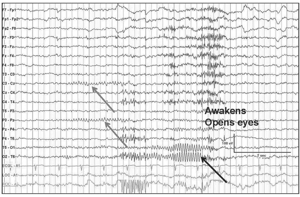

In some cases, it is not possible to determine the normality or abnormality unless a previous EEG is available for comparison. The normal low-voltage background pattern also tends to show well-defined and prominent photic driving responses with high-frequency photic stimulation. The measurable alpha rhythm may also appear during hyperventilation. In some normal people, the background activity is low voltage initially but may show measurable alpha rhythm in the latter portion of the recording. It doesnt show if theres any damage or physical abnormalities in your brain. 6 In some cases, it is difficult to differentiate if the low-voltage activity is abnormally Abnormal EEG Patterns correlation with underlying cerebral lesions and neurological diseases Suthida Yenjun Definition of the abnormal EEG An EEG is abnormal if it contains Epileptiform activity Slow waves Amplitude abnormalities or Deviations from normal patterns In most abnormal EEGs, the abnormal patterns appear only. An EEG test only gives information about the electrical activity in your brain. 5 There is some evidence that genetic factors play a role in determining the voltage pattern in a healthy person. The two main types of slowing are focal and generalized slowing. EEG can provide evidence for underlying diffuse or focal cerebral dysfunction through demonstration of background slowing.

Approximately 10% of normal subjects show a low-voltage (<20 µV) background pattern that is difficult to measure. The Abnormal EEG - Electroencephalography (EEG): An Introductory Text and Atlas of Normal and Abnormal Findings in Adults, Children, and Infants - NCBI Bookshelf.


 0 kommentar(er)
